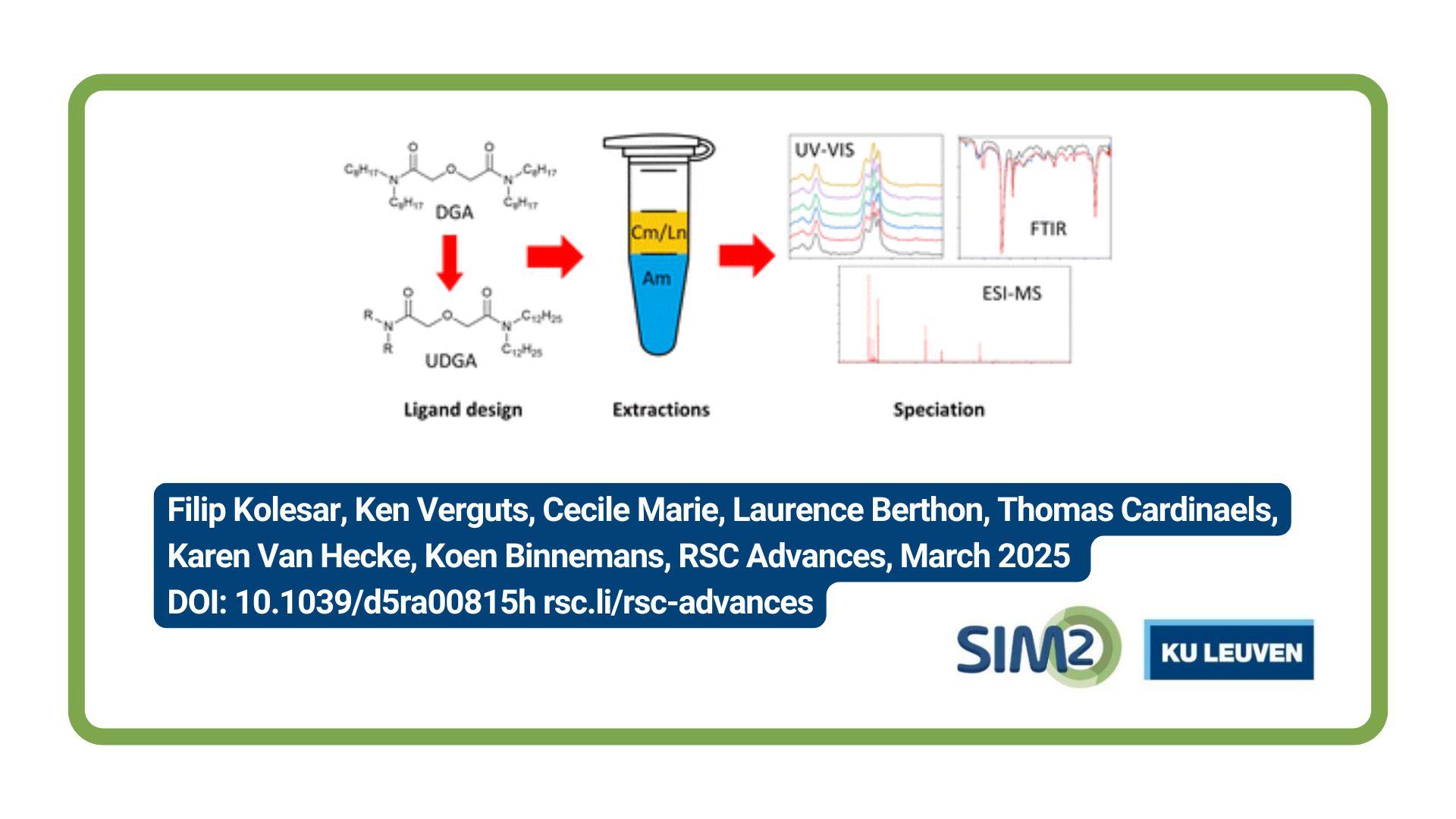

SIM² KU Leuven geologists Fernando Araujo, Niels Hulsbosch and Philippe Muchez have demonstrated that Raman spectroscopy is a powerful analytical tool to characterise and map complex mineral assemblages. Among others, the researchers focussed on phosphate minerals with light elements in their compositions, such as the lithium-bearing ore minerals montebrasite-amblygonite [LiAlPO4(OH,F)]. The work has been published in the Journal of Raman Spectroscopy.
Li-bearing phosphate minerals
Phosphate minerals, a group with more than 1800 species made of the phosphate [PO43-] oxyanion, are present in numerous geological and biological systems. As a mineral resource, phosphate rocks are classified as critical raw materials by the European Commission (CRM list 2020).
Phosphorus has its major use in chemical, pharmaceutical, and agricultural industries, but some phosphate minerals like montebrasite-amblygonite [LiAl(PO4)(OH,F)] are gaining economic interest as alternative lithium ores to cope with the increasing demand for Li-ion battery technology.
How to characterise?
Because montebrasite-amblygonite is a potential Li-ore, it is crucial to check samples for alteration products and to evaluate the presence of mineral impurities. In order to identify these minerals and understand their formation, a high spatial resolution mineralogical characterisation is required, as phosphates often co-occur with other minerals.
Moreover, many phosphates have complex compositions, and most analytical techniques (e.g. microprobe, SEM-EDS, etc.) are unable to directly measure some of its components, e.g. Li.
Consequently, phosphate mineral characterisation by most conventional quantitative (micro)analyses is challenging as these techniques either depend on X-ray chemical analyses or have low spatial resolution, and commonly require extensive sample preparation before analysis.
Confocal micro-Raman analysis, in turn, provides in situ molecular information and can overcome those restrictions. Additionally, the current technological advances in the field enable the fast acquisition of hyperspectral images, allowing the identification of different minerals and the visualisation of their spatial distributions.
Buranga pegmatite assessed by Raman spectroscopy
The study investigates two samples from the Buranga pegmatite, a P–Li–Nb–Ta-rich magmatic rock from Rwanda. The first case shows the replacement of primary F-rich montebrasite [LiAl(PO4)(OH,F)] by secondary F-poor montebrasite [LiAl(PO4)OH]. The second case looks into a complex sequence of phosphate minerals.
Raman mapping shows that higher temperature minerals are transformed into intermediate temperature phases and then to low-temperature minerals. Raman analyses can distinguish all the involved minerals with higher accuracy than regular optical microscopy or even electron microscopy.
By combining the information from both cases, a general trend of progressive hydration, followed by internal remobilisation of other elements, such as Na and Ca, can be inferred during cooling of the Buranga pegmatite.
In conclusion, Raman mapping is shown to be a powerful microanalytical technique to investigate complex mineral assemblages and their formation sequence. It can identify major and accessory phases and image their spatial relations in situ with high resolution and certainty. Raman images can aid in the search of high-quality Li-ore samples as it easily shows preferential regions with high concentration of minerals bearing this metal.
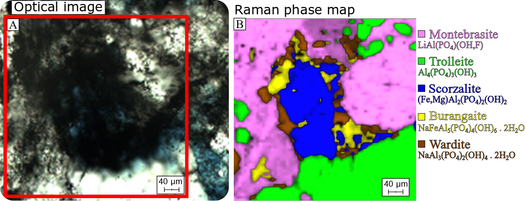
Figure 1. Raman mapping of a phosphatic mineral assemblage from the Buranga pegmatite, Rwanda. A) Optical photomicrograph under transmitted white light with a 50x objective and parallel polarisers. The red square marks the area of B. B) CLS multivariate statistical analysis output image from the mapping dataset showing the multiple minerals that are present in the mapped area. The legend on the right specifies the ideal chemical composition of each mineral identified.
Full reference of paper
Prado Araujo F, Hulsbosch N, Muchez P. High spatial resolution Raman mapping of complex mineral assemblages: Application on phosphate mineral sequences in pegmatites. J Raman Spectrosc. 2020;1–19. https:// doi.org/10.1002/jrs.6040
Acknowledgements
Fonds Wetenschappelijk Onderzoek, Grant/Award Number: I000718N; KU Leuven, Grant/Award Number: FLOF 000000132149
[credits featured image: Rob Lavinsky, iRocks.com – CC-BY-SA-3.0, CC BY-SA 3.0 <https://creativecommons.org/licenses/by-sa/3.0>, via Wikimedia Commons]